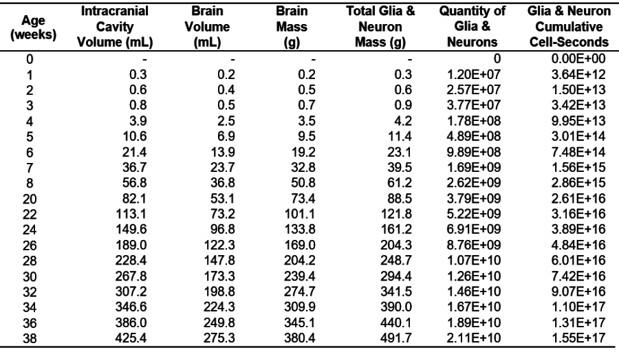
Human fetus growth model for glia and neurons from 0 to 38 weeks
References:
[1] Patrick, A.D. & Patrick, B.E., Carbon 14 Decay as a Source of Somatic Point Mutations in Genes Correlated with Cancer Diagnoses, Stable Isotope Foundation, Grants Pass, OR (2017).
[2] Luecke, R. H., Wosilait, W. D., and Young, J. F. Mathematical modeling of human embryonic and fetal growth rates, Growth Dev. Aging 63(1-2):49-59 (1999).
[3] Mehta S. Updated anatomical data and mathematical models for embryo/fetus dosimetry, Indian J. Nucl. Med. 27:101-4 (2012).
[4] Sender, R., Fuchs, S., & Milo, R. Revised estimates for the number of human and bacteria cells in the body, PLoS Biol 14(8): e1002533 (2016).
[5] Clouchoux C, Guizard N, Evans AC, et al. Normative fetal brain growth by quantitative in vivo magnetic resonance imaging, Am J Obstet Gynecol 206:173.e1-8 (2012).
[6] Dekaban, A.S. and Sadowsky, D., Changes in brain weights during the span of human life: relation of brain weights to body heights and body weights, Ann. Neurology 4:345-356 (1978).